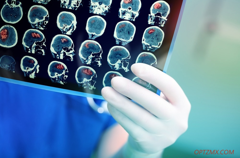
神经成像揭示了人脑的结构和功能。核磁共振成像、功能磁共振成像、CT、PET、脑电图等技术是发展我们对大脑如何工作以及在影响大脑的疾病中如何改变大脑的知识的基础,如神经组织退化性疾病(如阿尔茨海默病、帕金森病)。
这对于阐明大脑在心理健康障碍(例如抑郁症、焦虑症、成瘾、精神分裂症)中的工作方式至关重要。因此,神经影像学帮助我们创新了新的疾病治疗方法,从而为各种疾病的患者提供了更有效的治疗。 光学技术的发展正在影响神经成像平台的能力。光学的发展使神经成像领域受益匪浅,将其推向前所未有的高度,帮助其充分发挥潜力。在这里,我们讨论了如何在利用多模光纤发展的具体实例中实现这一点。我们思考了这将如何影响神经成像的未来结果,以及为什么这对整体健康很重要。 什么是多模光纤? 多模光纤是专门设计用于同时传输多种光线或光模式的光纤。这些光纤的光纤芯以微小不同的反射角传输每条光线或模式,从而可以同时传输多条光线或模式。 人们普遍认为,多模光纤是目前神经科学实验室必须与传统成像技术(如脑电图和功能磁共振成像)无法进入的大脑深层区域连接的最广泛的工具。 多模光纤如何用于神经成像? 多模光纤正在帮助增强当前的神经成像技术,使它们能够克服当前的限制,以便我们能够收集更可靠、更深入的人脑工作数据。有了这些数据,我们应该能够极大地扩展我们对人类大脑的理解,从而开发出治疗神经系统疾病的新方法。 2022 年发表在 APL Photonics 杂志上的一篇论文说明了如何利用多模光纤来改进成像技术,以可视化众所周知难以研究的大脑亚细胞结构。 微型内窥镜通常用于可视化大脑的深层结构。然而,这种方法的局限性在于它依赖于梯度指数 (GRIN) 微透镜,当它们被开发得更小时,它们会遭受越来越多的像差和受限的视野。这一主要限制使他们无法详细可视化大脑的深层结构,以揭示关键的结构和功能信息。 荷兰的研究人员通过在其内窥镜中利用多模光纤结合先进的波前工程技术克服了这一限制,由于高分辨率成像,使他们能够以最小的侵入性进入深层组织。这项研究是首次成功证明这一概念。 该团队通过单个 50μm芯多模光纤探头使用自发荧光人脑成像。该团队测试了两种方法:一种是通过光栅扫描成像获取图像的主动波前整形方法,另一种是使用基于散斑的压缩成像技术的计算图像恢复方法。 研究人员报告说,使用压缩成像的方法可以显着减少重要区域的图像采集时间,比传统技术允许的图像采集时间大三倍,同时保持高水平的空间分辨率。 他们的结果证明了脂褐质的积累,这是一种与阿尔茨海默病有关的与年龄有关的色素,捕获的可视化比传统方法快18倍。 为什么神经影像学很重要,它在未来将如何发展? 发表在 APL Photonics 上的研究中描述的多模光纤方法展示了一种对大脑深层结构进行成像的微创解决方案。 所提出的方法通过测量与单根头发纤维一样薄的设备提供了一种快速、高灵敏度和高分辨率的成像技术。 这种方法的适应可能对于开发敏感和有效的方法来成像精细和难以可视化的生物环境(例如深部脑组织、内脏和其他生物医学深层组织)至关重要。 其他神经影像学和临床研究领域可能会受益于这项技术的发展和以下进展。未来几年,这种方法可能会加速神经成像领域的发展,因为它提供了一种在难以研究的领域捕获数据的方法。 因此,我们可能会看到治疗神经组织退化性疾病(如阿尔茨海默病)的新方法的出现,并加深我们对精神健康疾病的神经相关性的理解。 How Multimode Optical Fibers Could Accelerate Neuroimaging Neuroimaging reveals the structure and function of the human brain. Techniques such as MRI, fMRI, CT, PET, EEG, and others have been fundamental in developing our knowledge of how the brain works and how it is altered in diseases that impact the brain, such as neurodegenerative disease (e.g. Alzheimer’s disease, Parkinson’s disease).
It has been vital in shedding light on how the brain works in mental health disorders (e.g., depression, anxiety, addiction, schizophrenia). As a result, neuroimaging has helped us to innovate novel ways to approach disease treatment, which has led to the more effective treatment of patients with a wide range of diseases. Developments in optical technology are influencing the capabilities of neuroimaging platforms. The evolution of optics is benefiting the field of neuroimaging, pushing it further than ever before to help it achieve its full potential. Here, we discuss how this is happening in the specific instance of leveraging developments in multimode optical fibers. We reflect on how this may influence future results in neuroimaging and why this is important to health overall. What are Multimode Optical Fibers? Multimode fibers are optical fibers specifically designed to transport multiple light rays or modes of light simultaneously. The optical fiber core of these fibers carries each ray or mode at a fractionally different reflection angle, making it possible to transport many simultaneously. It is generally accepted that multimode optical fibers are currently the most diffused tool that neuroscience laboratories have to interface with the brain’s deeper regions inaccessible to conventional imaging techniques such as EEG and fMRI. Related Stories · Noninvasive Neuroimaging Helps Recognize Culprit Behind Medication-Resistant Epilepsy · Photonic Crystal Fibers - Paving the Way for Better Optical Fibers · New Wearable Headset for Noninvasive Optical Neuroimaging While multimode optical fibers are often considered very limited systems, recent neuroimaging studies have revealed that advances in optics and photonics can be leveraged into neuroscience research, overcoming the limitations of traditional systems with multimode optical fibers. How are Multimode Optical Fibers Being Used in Neuroimaging? Multimode optical fibers are helping to enhance current neuroimaging techniques, allowing them to overcome current limitations so that we can gather more reliable, in-depth data on the workings of the human brain. With this data, we should be able to vastly expand our understanding of the human brain, and, therefore, develop novel approaches to treating neurological diseases. A paper published in the journal APL Photonics in 2022 illustrates how multimode optical fibers are being leveraged to improve imaging techniques employed to visualize the brain’s subcellular structures that are notoriously difficult to study. Miniaturized micro endoscopes are typically utilized to visualize the brain's deep structures. However, this method is limited in that it relies on gradient index (GRIN) microlenses that suffer from increasing aberrations and restricted fields of view as they are developed to become smaller and less invasive. This major limitation prevents them from visualizing the brain’s deep structures in detail necessary to reveal critical structural and functional information. Researchers in the Netherlands overcame this limitation by utilizing multimode optical fibers within their endoscopes combined with advanced wavefront engineering techniques, allowing them access to deep tissue with minimal invasiveness due to high-resolution imaging. This research is the first time this concept was successfully demonstrated. The team used auto-fluorescence human brain imaging via a single 50 μm-core multimode optical fiber probe. The team tested two approaches: an active wavefront shaping approach that acquired images by raster-scan imaging and a computational image recovery approach that used speckle-based compressive imaging technology. Advancing Brain Imaging with Two-Photon Fluorescence Microscopy The researchers reported that the approach using compressive imaging resulted in a significant decrease in image acquisition time for an area of interest up to three times larger than conventional techniques allow while maintaining high levels of spatial resolution. Their results demonstrated lipofuscin accumulation, the age-related pigment implicated in Alzheimer’s disease. The visualization captured was 18 times faster than conventional methods. Why is Neuroimaging Important and How Will it Evolve in the Future? The multimode optical fiber method described in the research published in APL Photonics demonstrates a minimally invasive solution to imaging deep brain structures. The proposed method offers a rapid, highly sensitive, and high-resolution imaging technique via apparatus that measures as thin as a single hair fiber. The adaptation of this method will likely be vital in developing sensitive and effective ways of imaging delicate and difficult-to-visualize biological surroundings, such as deep brain tissue, internal organs, and other biomedical deep tissues. Other neuroimaging and clinical research fields will likely benefit from this technique's development and the following advances. It is possible that, in the coming years, this method will accelerate developments in the field of neuroimaging, as it offers a way to capture data on hard-to-study areas. As a result, we may see the emergence of novel approaches to treating neurodegenerative diseases, such as Alzheimer’s disease, and growing our understanding of the neural correlates of mental health illnesses. References and Further Reading De Vittorio, M. and Pisanello, F., 2021. Multimode Optical Fibers for Optical Neural Interfaces. Advances in Experimental Medicine and Biology, pp.565-583. https://pubmed.ncbi.nlm.nih.gov/33398843/ Lochocki, B., Verweg, M., Hoozemans, J., de Boer, J. and Amitonova, L., 2022. Epi-fluorescence imaging of the human brain through a multimode fiber. APL Photonics, 7(7), p.071301. https://aip.scitation.org/doi/10.1063/5.0080672 Turtaev, S., Leite, I., Altwegg-Boussac, T., Pakan, J., Rochefort, N. and Čižmár, T., 2018. High-fidelity multimode fibre-based endoscopy for deep brain in vivo imaging. Light: Science & Applications, 7(1). https://www.nature.com/articles/s41377-018-0094-x Disclaimer: The views expressed here are those of the author expressed in their private capacity and do not necessarily represent the views of AZoM.com Limited T/A AZoNetwork the owner and operator of this website. This disclaimer forms part of the Terms and conditions of use of this website.
Written by After studying Psychology and then Neuroscience, Sarah quickly found her enjoyment for researching and writing research papers; turning to a passion to connect ideas with people through writing. |
联系我们|本论坛只支持PC端注册|手机版|小黑屋|吾爱光设 ( 粤ICP备15067533号 )
GMT+8, 2025-7-16 04:17 , Processed in 0.062500 second(s), 17 queries .
Powered by Discuz! X3.5
© 2001-2024 Discuz! Team.